Our services
Pulmonary Diagnosis
& Treatment Services
Better Breathing For a Better You.
Over 40 years of expert care in pulmonary,
sleep, and critical care medicine — across five convenient locations in Gwinnett County.


Rating 4.8/5.0
Need help with Sleeping Better instead?
Helping You Breathe Easier
We diagnose and treat a wide range of respiratory conditions with advanced tools and customized care plans.
Pulmonary Services We Offer:


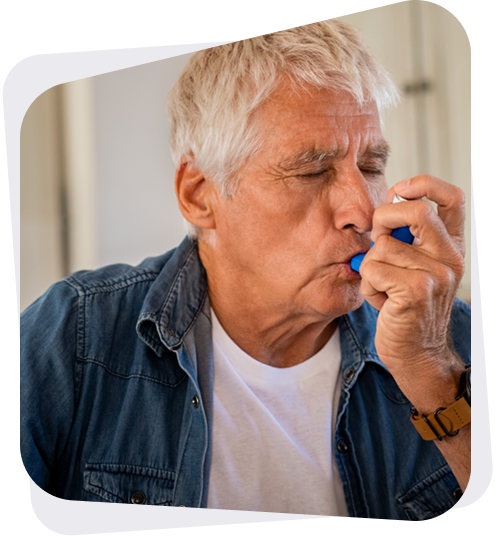
Trouble breathing or chronic cough?
All services available at our 5 convenient locations. No referral required for new patients.
More Ways to Heal
Comprehensive care that supports healing and enhances your comfort and recovery.
More Pulmonary Services Include:

Comprehensive Lung Care.
Our pulmonary program makes it easy to get expert respiratory
care close to home. From diagnostic testing to ongoing treatment
plans, our team helps you breathe easier with confidence.
- Pulmonary testing
- Lung evaluations
- Advanced bronchoscopies
- Chronic management
- Procedure follow-ups
- Patient education
Sleep and breathing news,made simple. Subscribe today.
Not Sure Where to Start? Here’s a quick guide:
Concern
Start Here
Shortness of breath
May indicate asthma, COPD, or restricted airflow—schedule testing to determine the cause.
Loud snoring or sleep apnea
Interrupted breathing at night? You may need a sleep study and CPAP therapy.
Chronic cough or lung nodules
Lingering cough or abnormal lung findings? Early evaluation is key to prevention and treatment.
Trouble sleeping or insomnia
Insomnia or fragmented sleep could signal a treatable sleep disorder. Let’s find out why.
Can’t tolerate CPAP?
Inspire is a surgical, mask-free option for sleep apnea patients who can’t use CPAP.
More Ways to Heal
Expert Care for Common Lung & Sleep Conditions
Helping You Breathe Easier
We diagnose and treat a wide range of
respiratory conditions with advanced
tools and customized care plans.
Diagnostic & Procedural Care
001
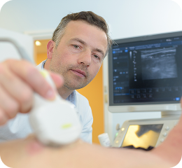
Pulmonary Ultrasound
Pulmonary ultrasound is a safe, non-invasive imaging tool that allows pulmonologists to evaluate the lungs and surrounding structures in real time. Using sound waves instead of radiation, this technology provides immediate insight into a variety of lung and chest conditions.
At Gwinnett Pulmonary & Sleep, pulmonary ultrasound plays an important role in diagnosing breathing concerns, detecting fluid around the lungs, and guiding certain procedures with precision. The exam is quick, painless, and often performed right at the bedside or in the office.
When accurate answers are needed without unnecessary delays or radiation exposure, pulmonary ultrasound is an effective and reliable option.
Book an Appointment | Contact Us
What Is Pulmonary Ultrasound?
Pulmonary ultrasound is a diagnostic imaging test that uses high-frequency sound waves to create detailed images of the lungs and pleural space (the area surrounding the lungs). A small handheld device called a transducer sends sound waves into the chest. These sound waves are reflected and converted into images displayed on a screen.
Unlike X-rays or CT scans, pulmonary ultrasound does not use radiation. The images are generated instantly, allowing your provider to evaluate lung movement, detect fluid, and assess abnormalities in real time.
This technology has become an important tool in modern pulmonary care because it is:
- Non-invasive
- Radiation-free
- Portable
- Immediate
Pulmonary ultrasound is especially valuable in situations where fast decision-making is important.
What Is Pulmonary Ultrasound Used For?
Pulmonary ultrasound can help evaluate a wide range of respiratory conditions. It is often used to clarify findings from physical exams or other imaging studies and can sometimes reduce the need for more extensive testing.
Common uses include:
Detecting Pleural Effusion (Fluid Around the Lungs)
One of the most common indications for a pulmonary ultrasound is to identify pleural effusion, a buildup of fluid between the lungs and chest wall. Ultrasound allows precise visualization of fluid and helps determine how much is present.
Evaluating Shortness of Breath
When someone experiences difficulty breathing, a pulmonary ultrasound can help determine whether the cause is fluid overload, infection, lung collapse, or another condition.
Identifying Pneumothorax (Collapsed Lung)
Pulmonary ultrasound is highly effective at detecting pneumothorax, a condition in which air collects outside the lung, causing it to collapse. Early detection allows for faster treatment.
Assessing Lung Infections
Ultrasound can reveal signs of pneumonia and other infections by identifying areas of lung consolidation or inflammation.
Monitoring Chronic Lung Conditions
For patients with ongoing respiratory disease, pulmonary ultrasound can help monitor changes over time without repeated radiation exposure.
Guiding Procedures
Pulmonary ultrasound is frequently used to guide procedures such as thoracentesis (removal of fluid from around the lungs). Real-time imaging improves accuracy and reduces the risk of complications.
Book an Appointment | Contact Us
How Pulmonary Ultrasound Works
Pulmonary ultrasound is straightforward and typically requires little to no preparation.
Before the Exam
In most cases, no special preparation is necessary. You can eat, drink, and take medications as instructed unless your provider advises otherwise.
The exam may be performed in-office or at the bedside for hospitalized patients:
Because the equipment is portable, pulmonary ultrasound can be performed wherever it is needed.
During the Exam
The procedure itself is simple and comfortable.
- You will sit upright or lie down, depending on the area being examined.
- A small amount of gel is applied to the skin. This gel helps the sound waves travel efficiently.
- The provider moves the handheld transducer across the chest wall.
- Images appear instantly on a monitor.
There are no needles involved unless the ultrasound is being used to guide a separate procedure. The exam typically takes about 15 to 30 minutes.
Pulmonary ultrasound does not cause pain. You may feel light pressure from the transducer, but the process is gentle.
After the Exam
There is no recovery time required. You can return to normal activities immediately after the exam.
Because images are available in real time, your provider may review findings with you shortly after the test is completed. In some cases, the results help guide immediate next steps in your care plan.
Benefits of Pulmonary Ultrasound
Pulmonary ultrasound offers several important advantages that make it a valuable tool in respiratory care.
No Radiation Exposure
Unlike CT scans and chest X-rays, pulmonary ultrasound uses sound waves rather than radiation. This makes it safe for repeated use and long-term monitoring.
Real-Time Imaging
Images are produced instantly, allowing providers to assess lung movement and fluid levels as they occur.
Non-Invasive and Comfortable
The exam does not require incisions, injections, or sedation.
Portable and Accessible
Because the equipment is compact and mobile, pulmonary ultrasound can be performed in various settings, including at the bedside for hospitalized patients.
Improved Safety During Procedures
When guiding fluid removal or other procedures, pulmonary ultrasound improves precision and reduces the risk of complications.
Faster Decision-Making
Immediate imaging supports faster diagnoses and timely treatment planning.
Is Pulmonary Ultrasound Safe?
Pulmonary ultrasound is considered extremely safe. It uses sound waves, a technology widely used in medical imaging for decades. There are no known harmful effects from diagnostic ultrasound when performed by trained professionals.
Because it does not involve radiation exposure, pulmonary ultrasound is appropriate for:
- Ongoing monitoring
- Repeated evaluations
- Patients of various ages
Safety and accuracy are priorities at Gwinnett Pulmonary & Sleep, and pulmonary ultrasound supports both.
How Pulmonary Ultrasound Compares to Other Imaging Tests
Pulmonary ultrasound does not completely replace chest X-rays or CT scans. Each imaging method has a specific role in pulmonary medicine.
Ultrasound is particularly effective for:
- Detecting fluid
- Assessing lung movement
- Identifying pneumothorax
- Guiding procedures
CT scans may still be necessary for detailed evaluation of lung nodules, tumors, or complex anatomy.
In many cases, pulmonary ultrasound serves as a first-line or complementary imaging modality that enhances diagnostic clarity without additional radiation exposure.
Why Choose Gwinnett Pulmonary & Sleep?
Advanced diagnostic tools are most effective when paired with experienced physicians and compassionate care.
Skilled Pulmonology Team
Our board-certified pulmonologists are trained in modern imaging techniques, including pulmonary ultrasound, to support accurate and efficient diagnosis.
Integrated Pulmonary Care
Pulmonary ultrasound is part of a comprehensive approach to lung health. When additional testing or treatment is needed, care is coordinated seamlessly.
Community-Focused Care
Gwinnett Pulmonary & Sleep proudly serves patients throughout Gwinnett County and areas north of Atlanta. With convenient locations and a dedicated team, expert pulmonary care is close to home.
Commitment to Patient Comfort
Clear communication, thoughtful guidance, and personalized care are central to every visit.
Schedule an Appointment
Pulmonary ultrasound is a safe, efficient method for evaluating lung conditions and guiding treatment decisions. Whether assessing fluid around the lungs, investigating shortness of breath, or supporting a procedure, this advanced imaging tool provides valuable insights without radiation exposure.
To learn more or schedule an appointment, contact Gwinnett Pulmonary & Sleep today. Our team is committed to delivering precise, compassionate pulmonary care for patients throughout Gwinnett County and north of Atlanta.
Book an Appointment | Contact Us
Frequently Asked Questions
How long does a pulmonary ultrasound take?
The exam typically lasts between 15 and 30 minutes.
Is pulmonary ultrasound painful?
No. The test is non-invasive and does not cause pain.
Does pulmonary ultrasound replace a CT scan?
Not always. While pulmonary ultrasound is highly effective for certain conditions, CT scans may still be necessary in some situations.
Are results available immediately?
Images are available in real time, and your provider may discuss findings during or shortly after the exam.
Is preparation required?
Most patients do not need special preparation before a pulmonary ultrasound.

001
Bronchoscopy
Pulmonary function tests (PFTs) measure how well your lungs take in and exhale air and how efficiently they transfer oxygen into the blood. There are several different tests. Spirometry measures how well the lungs exhale (breathe out). Lung volume measures how well the lungs inhale (breathe in). Testing the diffusion capacity of carbon monoxide (DLCO) shows how efficiently the lungs transfer oxygen from the air into the bloodstream.
Why are these tests done?
Pulmonary function tests help:
- Diagnose lung diseases such as asthma, chronic bronchitis, and emphysema.
- Determine the cause of shortness of breath.
- Measure the effects of exposure to chemicals, coal dust and other toxins on your lung function.
- Measure the effectiveness of medicines and other treatments.
- Detect lung disease at an early stage before you have symptoms.
How do I prepare for these tests?
Eat a light meal and do not smoke for four to six hours before your test. If you have asthma, ask your health care provider if you need to stop using asthma medicine before the test.
How is the test done?
Spirometry—You breathe into a mouthpiece connected to an instrument called a spirometer. The spirometer measures the volume of air that you can force out of your lungs in one second after having inhaled as much as you can. You will be asked to hold the tube of a spirometer in your mouth, inhale as much air as possible, and then blow out as hard as you can into the spirometer for one second. The amount of air you can force out is called your forced expiratory volume, or FEV1.
Lung volume: You breathe nitrogen or helium gas through a tube for a certain amount of time, and then the concentration of the gas in a chamber attached to the tube is measured.
Diffusion capacity—You breathe carbon monoxide for a very short time (often one breath). The concentration of carbon monoxide in the air you exhale is then measured. The difference in the amounts of carbon monoxide inhaled and exhaled shows how quickly gas can travel from your lungs into the blood.
PFTs are painless, and you will have time to rest between the different breathing measurements. The measurements may be repeated two or more times.
Pulse Oximetry
A pulse oximeter is a medical device that indirectly measures the oxygen saturation of a patient’s blood (as opposed to measuring oxygen saturation directly through a blood sample) and changes in blood volume in the skin, producing a photoplethysmograph. It is often attached to a medical monitor so staff can always see a patient’s oxygenation. Most monitors also display the heart rate.
A blood-oxygen monitor displays the percentage of arterial hemoglobin in the oxyhemoglobin configuration. Acceptable normal ranges are 95 to 100 percent, although values down to 90 percent are common. For a patient breathing room air, at not far above sea level, an arterial pO2 can be estimated from the blood-oxygen monitor SpO2 reading.
We also perform exercise pulse oximetry for Medicare patients needing to re-qualify for their home oxygen and evaluate a patient’s oxygen levels during exercise.
Coumadin Clinic
We offer an in-office Coumadin clinic for patients using Coumadin, Warfarin, Lovenox, and Fragmin. We perform a finger-stick PT/INR level check and consult with an RN.
What is the prothrombin time test?
The prothrombin time, or PT, test measures when it takes blood to form a clot. This test is also often called protime.
The results of the prothrombin time test may vary from lab to lab, so healthcare providers use a ratio called the INR (international normalized ratio) to be able to account for the differences.
Why is this test done?
The PT/INR is usually done to measure the effect of blood-thinning medicines (anticoagulants), such as Warfarin (Coumadin).
If you have a medical condition such as atrial fibrillation or deep vein thrombosis or have had a heart valve replaced, your blood is more likely to form clots. Clots can block blood vessels and possibly cause a heart attack or stroke. Your healthcare provider may prescribe a blood thinner to help prevent clots. It’s very important to measure the effect of a blood thinner with this test. The medicine should keep the blood just thin enough to prevent clots. If the blood is too thin, you may bleed too easily.
The prothrombin time test may also be done if you have abnormal bleeding or clotting.
Injections
At the Gwinnett Pulmonary Group, we offer a number of injections and vaccines meant to keep patients healthy at all times. These injections include:
- XOLAIR
- Used to treat patients with severe asthma
- VACCINES
- Pneumonia
- Flu
- H1N1
003
Pulmonary Clearance
When a surgery or specific job requires confirmation that your lungs are functioning properly, a comprehensive pulmonary clearance evaluation may be necessary. This assessment helps determine whether your respiratory health is stable and whether additional precautions or treatment adjustments are needed.
Pulmonary clearance plays an important role in reducing risk and ensuring safety before anesthesia, surgery, or employment in physically demanding roles. At Gwinnett Pulmonary & Sleep, our board-certified pulmonologists provide thorough evaluations designed to deliver clear documentation and coordinated recommendations.
Accurate assessment supports better outcomes and peace of mind.
Book an Appointment | Contact Us
What Is Pulmonary Clearance?
Pulmonary clearance is a medical evaluation of your lung health to determine whether you are fit to undergo surgery or meet employment-related respiratory requirements.
Pulmonary clearance focuses on identifying:
- Existing lung conditions
- Breathing limitations
- Risk factors related to anesthesia
- Potential complications during or after surgery
This evaluation may include a review of medical history, a physical exam, lung function testing, and imaging when appropriate.
Pulmonary clearance does not automatically prevent a procedure or employment opportunity. Instead, it helps guide informed decision-making and ensures that any risks are properly addressed in advance.
Pulmonary Clearance for Surgery
Pulmonary clearance for surgery is commonly requested before procedures that require anesthesia or carry respiratory risk.
Surgery and anesthesia can temporarily affect lung function. Patients with preexisting conditions such as COPD, asthma, pulmonary fibrosis, or a history of smoking may have a higher risk of postoperative breathing complications.
A pulmonary clearance for surgery evaluation helps:
- Assess baseline lung function
- Identify uncontrolled respiratory conditions
- Recommend optimization strategies before surgery
- Reduce the risk of postoperative pneumonia or respiratory failure
- Coordinate care with the surgical team
In some cases, adjustments to medications, inhalers, or oxygen therapy may be recommended prior to surgery. When needed, additional testing, such as pulmonary function tests or imaging, can provide more detailed information.
Clear communication between your pulmonologist and surgeon ensures that your procedure is planned with safety in mind.
Book an Appointment | Contact Us
Pulmonary Clearance for Employment
Certain occupations require verification of adequate lung function before hiring or returning to work. Pulmonary clearance for employment ensures that an individual can safely perform job duties that may involve physical exertion, exposure to respiratory irritants, or safety-sensitive tasks.
Examples of positions that may require pulmonary clearance include:
- Firefighters
- Law enforcement officers
- Industrial or factory workers
- Healthcare workers
- Commercial drivers
- Workers exposed to airborne particles or chemicals
The evaluation helps confirm that lung function meets occupational standards and that respiratory conditions are appropriately managed.
Pulmonary clearance for employment also protects workers by identifying potential health risks before exposure to demanding environments.
Book an Appointment | Contact Us
What to Expect During a Pulmonary Clearance Evaluation
A comprehensive pulmonary clearance appointment is structured and thorough.
Medical History Review
Your provider will review:
- Prior lung diagnoses
- Smoking history
- Medication use
- Past surgeries
- History of hospitalizations
- Current symptoms, such as shortness of breath or a chronic cough
Understanding your baseline health helps guide further testing.
Physical Examination
A focused physical exam evaluates breathing patterns, oxygen levels, and overall respiratory stability.
Pulmonary Function Testing
Pulmonary function tests (PFTs) measure how well your lungs move air in and out. These tests help identify:
- Airflow obstruction
- Restrictive lung disease
- Reduced lung capacity
- Gas exchange abnormalities
PFT results are often central to pulmonary clearance for surgery and employment decisions.
Imaging Studies
Chest X-rays or CT scans may be reviewed if needed to assess lung structure, nodules, scarring, or other abnormalities.
Additional Testing
Depending on the situation, additional evaluations may include:
- Oxygen testing
- Six-minute walk test
- Sleep study (if sleep apnea is suspected)
- Blood work
Each pulmonary clearance evaluation is individualized based on your health status and the requirements of the requesting surgeon or employer.
How to Get Pulmonary Clearance
Many patients ask how to get pulmonary clearance.
The process typically includes:
- Obtaining a referral from your surgeon or employer, if required.
- Scheduling an appointment with a pulmonologist.
- Bringing relevant medical records or test results.
- Completing any recommended testing.
Once the evaluation is complete, documentation is prepared and shared with the requesting provider or organization.
Timelines vary depending on whether additional testing is needed. In many cases, evaluations can be completed promptly to avoid delaying surgery or employment.
Where to Get Pulmonary Clearance
Pulmonary clearance is best performed by a board-certified pulmonologist with expertise in respiratory risk assessment.
A specialized pulmonary clinic offers:
- Advanced diagnostic equipment
- Comprehensive lung testing
- Experienced interpretation of results
- Clear communication with surgeons and employers
Gwinnett Pulmonary & Sleep provides pulmonary clearance services at multiple convenient locations throughout Gwinnett County and areas north of Atlanta.
Choosing experienced pulmonary specialists ensures that evaluations are accurate, thorough, and efficiently documented.
Book an Appointment | Contact Us
Why Choose Gwinnett Pulmonary & Sleep?
Pulmonary clearance requires both clinical expertise and efficient coordination.
Board-Certified Pulmonologists
Our physicians specialize in diagnosing and managing complex respiratory conditions.
Comprehensive Testing Capabilities
On-site coordination of pulmonary function testing and imaging streamlines the evaluation process.
Efficient Documentation
Clear and timely reporting helps prevent unnecessary delays in surgery or employment.
Coordinated Communication
We work directly with surgeons, primary care physicians, and employers, as needed, to ensure continuity of care.
Local, Accessible Care
Serving patients throughout Gwinnett County and north Atlanta, our practice offers convenient access to specialized pulmonary expertise.
Schedule Your Pulmonary Clearance Evaluation
If you need pulmonary clearance for surgery or employment, a timely evaluation helps ensure safety and prevent delays.
At Gwinnett Pulmonary & Sleep, comprehensive respiratory assessments are performed with precision and professionalism. Our team provides clear documentation and coordinated care to support your next steps.
Contact our office today to schedule your pulmonary clearance appointment. Proper preparation today supports safer procedures and confident progress forward.
Book an Appointment | Contact Us
Frequently Asked Questions
How long does pulmonary clearance take?
The appointment typically lasts 30 to 60 minutes, depending on required testing.
Is pulmonary clearance the same as a routine physical?
No. Pulmonary clearance specifically evaluates lung health and respiratory risk rather than general health status.
What happens if concerns are identified?
If issues are detected, your pulmonologist may recommend treatment adjustments or additional monitoring before proceeding with surgery or employment.
How long is pulmonary clearance valid?
Validity depends on the requirements of the surgeon or employer and whether your health status changes.
Do I need to stop smoking before surgery?
Smoking cessation is strongly recommended before surgery to reduce complications. Your provider can offer guidance and support.
003
PleurX Catheter Placement
Recurrent fluid buildup around the lungs can make breathing uncomfortable and limit daily activities. When pleural effusion continues to return after treatment, a longer-term solution may be recommended. PleurX catheter placement is a minimally invasive procedure that allows fluid to be safely drained at home, helping improve breathing and reduce the need for repeated office visits.
At Gwinnett Pulmonary & Sleep, we perform chest PleurX catheter placement with precision and careful guidance. This approach offers ongoing symptom relief while supporting comfort, independence, and quality of life.
Book an Appointment | Contact Us
What Is a PleurX Catheter?
A PleurX catheter is a small, flexible tube placed into the pleural space, the area between the lungs and the chest wall, where excess fluid can accumulate.
This procedure involves inserting a soft drainage tube into the chest. Once in place, the catheter allows fluid to be removed safely and gradually at home using special drainage bottles.
This option is often recommended for patients who experience recurrent pleural effusion, meaning fluid continues to build up despite previous treatments such as thoracentesis.
Unlike a one-time drainage procedure, the placement of a PleurX catheter provides ongoing access for fluid removal without the need for repeated needle insertions.
Why PleurX Catheter Placement Is Performed
Fluid accumulation around the lungs can occur for several reasons, including:
- Certain cancers
- Heart failure
- Chronic lung conditions
- Liver or kidney disease
- Inflammatory disorders
When fluid repeatedly returns, it can cause:
- Shortness of breath
- Chest pressure
- Persistent cough
- Fatigue
Repeated thoracentesis procedures may temporarily relieve symptoms, but if fluid continues to reaccumulate, a more sustainable solution is often needed.
PleurX catheter placement is designed to:
- Provide consistent symptom relief
- Reduce the need for frequent procedures
- Allow drainage at home
- Improve comfort and breathing
For many patients, this option reduces hospital visits and allows more control over symptom management.
Book an Appointment | Contact Us
The PleurX Catheter Placement Procedure
The PleurX catheter placement procedure is typically performed as an outpatient procedure using imaging guidance to ensure accuracy and safety.
Before the Procedure
Prior to chest PleurX catheter placement, your provider will:
- Review imaging studies, such as chest X-ray or CT scan
- Confirm the presence and location of fluid
- Review medications, especially blood thinners
- Provide instructions about preparation
Ultrasound is commonly used to precisely identify the fluid collection and guide catheter placement.
During the Procedure
The procedure is performed under local anesthesia to numb the area.
Here is what typically occurs during the placement of a PleurX catheter:
- The skin is cleaned and prepared.
- A small incision is made in the chest wall.
- Using ultrasound guidance, the catheter is gently inserted into the pleural space.
- The catheter is tunneled under the skin to reduce the risk of infection.
- The tube is secured and covered with a sterile dressing.
The PleurX catheter placement procedure usually takes about 30 to 60 minutes.
Patients may feel pressure during the process, but significant pain is uncommon due to local anesthesia.
After the Procedure
Following PleurX catheter placement, patients are monitored briefly before returning home.
You may experience:
- Mild soreness at the insertion site
- Temporary fatigue
- Slight discomfort when draining fluid initially
Most individuals resume light activities within a day or two, depending on overall health.
Your care team will provide detailed instructions on managing the catheter and performing drainage safely.
How the PleurX Catheter Works at Home
One of the primary benefits of PleurX catheter placement is the ability to manage fluid drainage at home.
The catheter connects to specially designed vacuum bottles that gently remove fluid from the pleural space. Drainage frequency varies based on how quickly fluid accumulates. Some patients drain fluid daily at first, then less often as production slows.
The process is designed to be simple and manageable. Patients or caregivers are trained on:
- How to connect the drainage bottle
- How to monitor fluid output
- How to keep the area clean
- When to contact the provider
In some cases, fluid production gradually decreases over time. When drainage is no longer needed, the catheter can be removed.
Benefits of PleurX Catheter Placement
PleurX catheter placement offers several advantages for patients experiencing recurrent pleural effusion.
Fewer Repeat Procedures
Instead of undergoing multiple thoracentesis procedures, the catheter allows ongoing drainage without repeated needle insertions.
Improved Breathing
Regular fluid removal reduces pressure on the lungs, making breathing easier.
Greater Independence
Drainage at home provides flexibility and reduces frequent clinic visits.
Outpatient Procedure
The chest PleurX catheter placement procedure is typically performed without the need for extended hospitalization.
Enhanced Comfort
Consistent fluid management helps reduce symptom flare-ups and improves overall quality of life.
Is PleurX Catheter Placement Safe?
The PleurX catheter placement procedure is considered safe when performed by experienced pulmonologists using imaging guidance.
Possible risks include:
- Infection
- Bleeding
- Catheter blockage
- Minor discomfort
Tunneling the catheter under the skin and using sterile techniques helps reduce the risk of infection. Regular follow-up appointments ensure the catheter continues to function properly.
At Gwinnett Pulmonary & Sleep, patient safety is always a priority. Each patient is carefully evaluated to determine whether a PleurX catheter placement is appropriate.
PleurX Catheter Placement vs Repeat Thoracentesis
Thoracentesis and PleurX catheter placement both remove fluid, but they serve different purposes.
- Thoracentesis is often a one-time or occasional drainage procedure.
- PleurX catheter placement provides a longer-term solution for recurring fluid buildup.
For patients with frequent fluid reaccumulation, the catheter may reduce the need for repeated procedures and improve comfort.
Your provider will recommend the most appropriate approach based on your condition and overall health.
Why Choose Gwinnett Pulmonary & Sleep?
Experience and coordinated care make a meaningful difference in procedural outcomes.
Advanced Pulmonary Expertise
Our board-certified pulmonologists have extensive experience performing chest procedures, including PleurX catheter placement.
Ultrasound-Guided Precision
Imaging guidance enhances safety and accuracy during placement.
Comprehensive Follow-Up
Ongoing monitoring ensures proper catheter function and promptly addresses any concerns.
Local, Accessible Care
Gwinnett Pulmonary & Sleep proudly serves patients throughout Gwinnett County and areas north of Atlanta, providing expert pulmonary care close to home.
Patient-Focused Approach
Clear education, hands-on training, and responsive support help patients feel confident managing their care.
Schedule a Consultation
If recurrent pleural effusion is affecting your breathing and comfort, placement of a PleurX catheter may provide a practical and effective solution.
At Gwinnett Pulmonary & Sleep, we combine advanced procedural expertise with compassionate care to support long-term symptom management.
Contact our office today to schedule a consultation and discuss whether a PleurX catheter placement procedure is right for you.
Book an Appointment | Contact Us
Frequently Asked Questions
How long does a PleurX catheter stay in place?
The catheter may remain in place for weeks or months, depending on fluid production. In some cases, it can be removed once fluid buildup decreases.
Is PleurX catheter placement painful?
Local anesthesia minimizes discomfort during the procedure. Mild soreness afterward is common but manageable.
Can I shower with a PleurX catheter?
Your care team will provide instructions on protecting the catheter site during bathing.
Who performs the drainage?
Patients or caregivers can perform drainage after receiving proper training. Home health services may also assist if needed.
Is PleurX catheter placement covered by insurance?
Coverage varies, but most insurance plans cover medically necessary catheter placement. Our office can help verify benefits.
003
In-Office Thoracentesis
Thoracentesis is a minimally invasive procedure used to remove excess fluid from the space around the lungs. When fluid builds up between the lung and the chest wall, it can cause shortness of breath, chest pressure, and discomfort. Removing that fluid can provide significant relief and help determine the underlying cause.
At Gwinnett Pulmonary & Sleep, thoracentesis can often be performed safely in the office under ultrasound guidance. This allows patients to receive timely care without hospital admission in many cases.
Prompt evaluation and treatment can improve breathing, clarify diagnosis, and support the next steps in your care. Appointments can be made within the same week, as scheduling allows.
Book an Appointment | Contact Us
What Is Thoracentesis?
Thoracentesis is a procedure that removes fluid from the pleural space, the thin area between the lungs and the chest wall.
The lungs normally sit within a small, lubricated space that allows them to expand and contract smoothly during breathing. When excess fluid accumulates in this area, the condition is called a pleural effusion.
Pleural effusion may occur due to:
- Heart failure
- Pneumonia or lung infection
- Cancer
- Inflammatory conditions
- Liver or kidney disease
The thoracentesis procedure removes this fluid using a small needle. The fluid may be sent to a laboratory for analysis to determine its cause.
Thoracentesis serves two important purposes:
- Diagnostic – to identify why fluid has developed
- Therapeutic – to relieve symptoms and improve breathing
Why Thoracentesis Is Performed
Fluid around the lungs can make it difficult to take a full breath. Even moderate amounts of fluid may cause:
- Shortness of breath
- Chest tightness
- Fatigue
- Persistent cough
Thoracentesis can quickly reduce these symptoms by removing excess fluid.
Diagnostic Evaluation
When the cause of fluid accumulation is unclear, thoracentesis allows the fluid to be analyzed in a laboratory. Testing may look for:
- Infection
- Cancer cells
- Signs of inflammation
- Protein levels
- Other chemical markers
Accurate diagnosis is essential in determining the appropriate treatment plan.
Symptom Relief
In addition to diagnosis, thoracentesis often provides noticeable improvement in breathing. Many patients experience relief shortly after fluid removal.
Book an Appointment | Contact Us
How the Thoracentesis Procedure Works
Thoracentesis is typically straightforward and well-tolerated.
Before the Procedure
Before performing thoracentesis, your provider will:
- Review recent imaging studies (such as chest X-ray or CT scan)
- Perform a physical exam
- Review medications, especially blood thinners
- Use ultrasound to locate the fluid precisely
Ultrasound guidance improves safety and accuracy by precisely localizing fluid accumulation.
During the Thoracentesis Procedure
The procedure is usually performed while you are sitting upright, leaning slightly forward with support.
Here is what typically happens:
- The skin on your back is cleaned thoroughly.
- A local anesthetic is used to numb the area.
- Using ultrasound guidance, a thin needle is inserted into the pleural space.
- Fluid is gently withdrawn through the needle into a collection container.
The thoracentesis procedure typically takes about 20 to 30 minutes.
You may feel mild pressure during fluid removal, but sharp pain is uncommon. If you experience discomfort, your provider can pause or adjust as needed.
After the Procedure
Once fluid removal is complete:
- The needle is removed
- A small bandage is placed over the site
- You are monitored briefly
In many cases, patients return home shortly after the procedure.
Mild soreness at the insertion site is possible. Most people resume normal activities within a day, unless otherwise instructed.
Your provider will review fluid test results once they are available and discuss next steps if needed.
Contact your provider promptly if you experience:
- Worsening shortness of breath
- Chest pain
- Fever
- Persistent coughing
Complications are uncommon, but monitoring ensures safety.
Benefits of In-Office Thoracentesis
Performing thoracentesis in the office offers several advantages:
Convenience
Avoiding hospital admission saves time and reduces stress.
Faster Care
Prompt scheduling, including same-week appointments, enables earlier symptom relief and more rapid diagnostic clarification.
Ultrasound Guidance
Real-time imaging increases accuracy and reduces the risk of complications.
Minimally Invasive
Thoracentesis does not require surgical incisions or general anesthesia.
Improved Comfort
Local anesthesia and careful technique help ensure a smooth experience.
Is Thoracentesis Safe?
Thoracentesis is considered a safe and commonly performed procedure when done by experienced clinicians.
Possible risks include:
- Minor bleeding
- Temporary cough
- Infection (rare)
- Pneumothorax (collapsed lung), which is uncommon and typically mild if it occurs
Ultrasound guidance significantly reduces the risk of complications. At Gwinnett Pulmonary & Sleep, patient safety is always a priority.
Thoracentesis vs Chest Drain
Thoracentesis is typically:
- A one-time or occasional procedure
- Used to remove fluid for diagnosis or symptom relief
A chest drain (or chest tube) is:
- A larger tube plis aced into the chest
- Used for continuous drainage over several days
- Often required in more severe or ongoing conditions
In many cases, thoracentesis provides effective relief without the need for longer-term drainage. Your provider will determine which option is appropriate based on your condition.
Why Choose Gwinnett Pulmonary & Sleep?
Choosing experienced specialists matters when undergoing any procedure.
Expertise in Pulmonary Care
Our board-certified pulmonologists are highly trained in performing thoracentesis safely and effectively.
Ultrasound-Guided Precision
Advanced imaging technology enhances accuracy and safety.
Comprehensive Evaluation
Thoracentesis is part of a broader pulmonary care plan that includes advanced diagnostics and follow-up.
Community-Based Care
Gwinnett Pulmonary & Sleep proudly serves patients throughout Gwinnett County and areas north of Atlanta, providing expert care close to home.
Patient-Focused Approach
Clear communication, thoughtful preparation, and attentive follow-up are central to every visit.
Schedule an Appointment
If you are experiencing shortness of breath or have been diagnosed with pleural effusion, thoracentesis may provide both relief and important diagnostic information.
At Gwinnett Pulmonary & Sleep, the thoracentesis procedure is performed with precision, safety, and patient comfort in mind. In-office care allows timely evaluation and treatment without unnecessary hospital visits.
Contact our office today to schedule a consultation and discuss whether thoracentesis is appropriate for your condition. Appointments may be made within the same week.
Book an Appointment | Contact Us
Frequently Asked Questions
How long does thoracentesis take?
The thoracentesis procedure usually takes 20 to 30 minutes.
Is thoracentesis painful?
Local anesthesia is used to numb the area. Most patients report mild pressure rather than significant pain.
How much fluid is removed?
The amount depends on the amount of fluid present. Your provider removes a safe volume based on your individual condition.
Will the fluid come back?
In some conditions, fluid may reaccumulate. Your provider will discuss long-term management if needed.
Can I drive afterward?
Depending on your overall health and comfort level, many patients are able to drive home. Your provider will give specific guidance.
003
Lung Nodule Risk Assessment
A lung nodule is a small spot or shadow that appears on a chest X-ray or CT scan. These nodules are common and are often discovered during imaging performed for another reason. While hearing that you have a lung nodule can feel concerning, most lung nodules are not cancerous.
A thorough lung nodule assessment helps determine whether a nodule is low risk and simply needs monitoring, or whether additional testing is necessary. At Gwinnett Pulmonary & Sleep, we take a structured, evidence-based approach to evaluating lung nodules so that each patient receives the right level of care, no more and no less.
Careful assessment provides clarity, direction, and peace of mind.
Book an Assessment | Contact Us
What Is a Lung Nodule?
A lung nodule is a small growth in the lung that is typically less than 3 centimeters in size. It may also be called a “spot on the lung” or a “pulmonary nodule.”
Lung nodules are frequently caused by:
- Old infections
- Scar tissue
- Inflammation
- Benign growths
In many cases, they are harmless. However, some nodules may represent early lung cancer, which is why a proper lung nodule assessment is important.
The purpose of evaluation is not to assume the worst; it is to accurately determine the level of risk and guide next steps appropriately.
What Is a Lung Nodule Assessment?
A lung nodule assessment is a comprehensive evaluation used to estimate the likelihood that a lung nodule is benign (non-cancerous) or malignant (cancerous).
This assessment does not rely on one single factor. Instead, it considers:
- Imaging characteristics
- Size and appearance of the nodule
- Growth over time
- Personal risk factors
Using established medical guidelines and clinical experience, pulmonologists determine whether the safest approach is monitoring, additional imaging, or biopsy.
Not every lung nodule requires invasive testing. In fact, many nodules can be safely observed over time.
Factors Considered During a Lung Nodule Assessment
A thorough lung nodule assessment involves careful review of several important elements.
Nodule Size
Size plays a significant role in risk evaluation. Smaller nodules are generally less likely to be cancerous, while larger nodules may require closer attention.
Your provider will measure the nodule precisely using CT imaging.
Shape and Appearance
The characteristics of the nodule provide valuable clues.
For example:
- Smooth, round nodules are often benign
- Irregular or spiculated edges may raise concern
- Solid nodules behave differently from ground-glass nodules
These imaging details are essential components of a structured lung nodule assessment.
Growth Over Time
Comparing current imaging with prior scans helps determine whether a nodule has changed.
A nodule that remains stable over time is often reassuring. A growing nodule may require further evaluation.
When previous scans are available, they provide valuable context for decision-making.
Personal Risk Factors
A lung nodule assessment also includes an evaluation of personal health history.
Important factors include:
- Age
- Smoking history
- Exposure to environmental toxins
- Family history of lung cancer
- History of prior cancers
Each person’s risk profile is unique, and recommendations are tailored accordingly.
What Happens After a Lung Nodule Assessment?
Once the evaluation is complete, your provider will recommend the most appropriate next step. The plan depends on your individual risk level.
Monitoring with Follow-Up Imaging
For low-risk nodules, observation is often the safest option.
This may involve:
- Scheduled CT scans at specific intervals
- Ongoing comparison of imaging results
- Continued monitoring over months or years
This approach avoids unnecessary procedures while maintaining vigilance.
Additional Imaging
If further clarification is needed, advanced imaging such as a PET scan may be recommended. PET scans can help assess metabolic activity within a nodule.
This step is often part of a more detailed lung nodule assessment when imaging findings are uncertain.
Biopsy or Diagnostic Procedures
If a nodule appears at higher risk, a biopsy may be necessary to obtain a tissue diagnosis.
Advanced diagnostic options may include:
- Robotic navigational bronchoscopy
- Endobronchial ultrasound (EBUS)
- CT-guided needle biopsy
At Gwinnett Pulmonary & Sleep, access to advanced, minimally invasive procedures enables accurate diagnosis when needed.
Book an Assessment | Contact Us
Why Early and Accurate Assessment Matters
Early evaluation supports better outcomes.
When lung cancer is detected at an early stage, treatment options are more effective and less invasive. At the same time, unnecessary procedures can be avoided for nodules that pose little risk.
A thoughtful assessment of a lung nodule balances caution with practicality.
Benefits of early evaluation include:
- Reduced anxiety through clear answers
- Timely treatment if necessary
- Avoidance of unnecessary surgery
- Evidence-based decision-making
Careful assessment ensures that each patient receives appropriate, personalized care.
Our Approach to Lung Nodule Assessment
At Gwinnett Pulmonary & Sleep, lung nodule evaluation is guided by established clinical guidelines and years of pulmonary expertise.
Evidence-Based Evaluation
Our pulmonologists follow nationally recognized guidelines to determine the appropriate level of monitoring or intervention. Each evaluation is a personalized process that begins with detection and diagnosis and follows the specific patient journey through treatment planning and follow-ups.
Advanced Diagnostic Capabilities
When further testing is needed, we offer advanced options such as:
- Robotic bronchoscopy
- Endobronchial ultrasound
- Pulmonary imaging and ultrasound
Access to these technologies enables seamless progression from assessment to diagnosis, if required.
Personalized Risk Stratification
Every lung nodule assessment is individualized. Recommendations are based on your imaging findings, medical history, and overall health profile.
Community-Focused Care
Gwinnett Pulmonary & Sleep proudly serves patients throughout Gwinnett County and areas north of Atlanta. Expert pulmonary evaluation is available close to home, with multiple convenient locations.
Schedule a Lung Nodule Assessment
If imaging identifies a lung nodule, a comprehensive assessment provides clarity and direction. Careful evaluation ensures that low-risk nodules are monitored appropriately while higher-risk findings are addressed promptly.
At Gwinnett Pulmonary & Sleep, advanced diagnostics and experienced pulmonologists work together to deliver accurate, personalized care.
To schedule a consultation or discuss your imaging results, contact our office today. Early evaluation supports informed decisions and confident next steps.
Book an Assessment | Contact Us
Frequently Asked Questions
Are lung nodules common?
Yes. Lung nodules are common, especially among individuals with a history of infections or prior lung inflammation.
Does a lung nodule mean cancer?
No. Most lung nodules are benign. A structured lung nodule assessment helps determine the risk level.
What size lung nodule is concerning?
Larger nodules may require closer monitoring or additional testing, but size alone does not determine risk.
How long does monitoring last?
Monitoring schedules vary depending on risk level. Some nodules require imaging over several months or years to confirm stability.
When is a biopsy necessary?
A biopsy may be recommended if imaging features or growth patterns suggest a higher likelihood of malignancy.
003
Endobronchial Ultrasound (EBUS)
When imaging tests show an abnormality in the lungs or surrounding lymph nodes, the next step is often finding out exactly what it means. At Gwinnett Pulmonary & Sleep, we use advanced diagnostic tools to provide clear answers while prioritizing your comfort and safety. One of the most important tools we offer is endobronchial ultrasound (EBUS).
EBUS is a minimally invasive procedure that allows our pulmonologists to examine the lungs and surrounding lymph nodes using ultrasound technology. By combining bronchoscopy with real-time imaging, we can obtain precise tissue samples without surgery in many cases.
Book an Appointment | Contact Us
What Is Endobronchial Ultrasound (EBUS)?
Endobronchial ultrasound is a specialized bronchoscopy technique that uses ultrasound imaging to visualize structures beyond the airways, including lymph nodes and masses.
During an endobronchial ultrasound bronchoscopy, a thin, flexible tube called a bronchoscope is gently guided through your mouth into your airways. Unlike traditional bronchoscopy, this scope incorporates a small ultrasound probe. The ultrasound produces real-time images of tissues and lymph nodes located just outside the airway walls.
This allows your physician to see areas that cannot be viewed with standard bronchoscopy alone.
Endobronchial Ultrasound vs Bronchoscopy: What’s the Difference?
A traditional bronchoscopy allows your doctor to look inside your airways to check for blockages, inflammation, tumors, or infection. However, it does not allow clear visualization of structures outside the airway walls.
With bronchoscopy with EBUS, ultrasound imaging is added. This makes it possible to:
- View lymph nodes near the lungs
- Identify abnormalities outside the airway
- Guide needle biopsies in real time
In simple terms, EBUS adds a “live ultrasound camera” to bronchoscopy, improving accuracy and reducing the need for more invasive procedures.
Why Is an EBUS Procedure Performed?
Your provider may recommend an EBUS procedure if imaging tests show abnormalities that require further evaluation.
Common reasons include:
Evaluating Enlarged Lymph Nodes
If lymph nodes in your chest appear enlarged on imaging, EBUS can help determine the cause.
Diagnosing or Staging Lung Cancer
Endobronchial ultrasound plays an important role in diagnosing lung cancer and determining whether it has spread to nearby lymph nodes. Accurate staging is essential for developing the right treatment plan.
Investigating Abnormal CT Scan Findings
If your CT scan shows a mass or area of concern, EBUS may help provide a tissue diagnosis without the need for surgical biopsy.
Diagnosing Infections or Inflammatory Conditions
Conditions such as sarcoidosis, tuberculosis, or certain fungal infections may require lymph node sampling for diagnosis. EBUS bronchoscopy allows us to collect these samples safely.
Book an Appointment | Contact Us
How the EBUS Procedure Works
Understanding what to expect can help ease concerns.
Before the Procedure
Before your endobronchial ultrasound bronchoscopy, your provider will:
- Review your imaging studies
- Go over your medications
- Provide instructions about fasting
- Discuss whether to adjust blood thinners
Our team will answer your questions and ensure you feel prepared.
During Bronchoscopy with Endobronchial Ultrasound
The procedure is typically performed in a hospital or specialized outpatient setting.
Sedation and Comfort
Most patients receive sedation or anesthesia so they remain comfortable throughout the procedure. You should not feel pain during the exam.
Real-Time Ultrasound Imaging
During bronchoscopy with EBUS, the bronchoscope is gently inserted through your mouth into your airways. The ultrasound probe produces live images of structures surrounding your lungs.
This real-time guidance allows your pulmonologist to precisely locate lymph nodes or masses.
Needle Biopsy
Once the targeted area is identified, a very fine needle is passed through the airway wall into the lymph node or mass to collect tissue samples. Because the ultrasound shows the exact location, the biopsy can be performed with high accuracy.
The entire EBUS procedure typically lasts about 30 to 60 minutes, depending on the complexity of the case.
After the Procedure
After your EBUS bronchoscopy, you will be monitored while the sedation wears off. Most patients go home the same day.
You may experience:
- Mild sore throat
- Hoarseness
- Light coughing
- Fatigue
These symptoms are usually temporary and improve within a day or two.
Your physician will let you know when to expect biopsy results, which are often available within several days.
Contact your provider immediately if you experience:
- Severe chest pain
- Difficulty breathing
- Fever
- Persistent bleeding
Complications are uncommon, but your safety is always our priority.
Benefits of Endobronchial Ultrasound
Endobronchial ultrasound offers several important advantages:
Minimally Invasive
Unlike surgical biopsy procedures, EBUS does not require external incisions. The procedure is performed through your natural airways.
Avoids Surgery in Many Cases
Because bronchoscopy with EBUS allows precise sampling of lymph nodes, many patients can avoid more invasive surgical procedures.
Real-Time Precision
The addition of ultrasound improves targeting accuracy and diagnostic confidence.
Faster Recovery
Most patients return home the same day and resume normal activities shortly afterward.
Important for Early Diagnosis
When evaluating possible lung cancer or other serious conditions, early and accurate diagnosis is critical. EBUS supports timely decision-making.
Is EBUS Safe?
Yes. EBUS bronchoscopy is considered a safe and well-established procedure when performed by experienced pulmonologists.
As with any medical procedure, there are potential risks, including:
- Minor bleeding
- Infection
- Temporary low oxygen levels
- Rare complications such as lung collapse
At Gwinnett Pulmonary & Sleep, we take every precaution to ensure your safety. Our team carefully evaluates each patient to determine whether endobronchial ultrasound is the right diagnostic approach.
Why Choose Gwinnett Pulmonary & Sleep for EBUS?
Choosing the right team for advanced diagnostic procedures matters.
Experienced Pulmonology Specialists
Our board-certified pulmonologists have extensive experience performing endobronchial ultrasound bronchoscopy and other advanced airway procedures.
Advanced Technology
We use modern, high-resolution ultrasound systems to improve diagnostic accuracy and patient safety.
Patient-Centered Approach
We understand that waiting for answers can be stressful. Our team is committed to:
- Clear communication
- Compassionate care
- Thorough follow-up
- Personalized treatment planning
We proudly serve patients throughout Gwinnett County and areas north of Atlanta, offering expert pulmonary care close to home.
Schedule Your Consultation
If you’ve been referred for further lung evaluation or need answers about abnormal imaging results, endobronchial ultrasound (EBUS) may provide the clarity you need.
At Gwinnett Pulmonary & Sleep, we combine advanced diagnostic technology with compassionate care to help you move forward with confidence.
Call our office today to schedule an appointment.
Or request a consultation online to learn whether an EBUS procedure is right for you.
Book an Appointment | Contact Us
Frequently Asked Questions
What is EBUS used for?
EBUS is used to evaluate enlarged lymph nodes, diagnose lung cancer, stage cancer, and investigate abnormal imaging findings.
Is EBUS the same as bronchoscopy?
No. Traditional bronchoscopy looks inside the airways. Bronchoscopy with endobronchial ultrasound adds ultrasound imaging to see structures outside the airways.
How long does the EBUS procedure take?
The procedure usually takes 30–60 minutes, depending on how many areas need evaluation.
Will I be asleep during EBUS?
Most patients receive sedation or anesthesia to ensure comfort.
When will I get results?
Biopsy results are typically available within a few days. Your provider will review them with you and discuss next steps.
Is EBUS painful?
You should not feel pain during the procedure. Mild throat irritation afterward is common but temporary.
Trouble breathing or chronic cough?
All services available at our 5 convenient locations. No referral required for new patients.
More Ways to Heal
Comprehensive care that supports healing and enhances your comfort and recovery.
Ongoing & Preventive Management
What kind of help do you need?
Comprehensive Lung Care.
Our pulmonary program makes it easy to get expert respiratory
care close to home. From diagnostic testing to ongoing treatment
plans, our team helps you breathe easier with confidence.
- Pulmonary testing
- Lung evaluations
- Advanced bronchoscopies
- Chronic management
- Procedure follow-ups
- Patient education
Sleep and breathing news,
made simple. Subscribe today.
Not Sure Where to Start? Here’s a quick guide:
Concern
Start Here
Shortness of breath
May indicate asthma, COPD, or restricted airflow—schedule testing to determine the cause.
Pulmonary Services
Loud snoring or sleep apnea
Interrupted breathing at night? You may need a sleep study and CPAP therapy.
Sleep Study or CPAP
Chronic cough or lung nodules
Lingering cough or abnormal lung findings? Early evaluation is key to prevention and treatment.
Pulmonary Evaluation
Trouble sleeping or insomnia
Insomnia or fragmented sleep could signal a treatable sleep disorder. Let’s find out why.
Sleep Medicine
Contact & Scheduling
We're here when you need us —
and we're accepting new patients.

Gwinnett Pulmonary & Sleep: 770.995.0630
Gwinnett Pulmonary & Sleep: 770.995.0630
Gwinnett Sleep: 678.942.5982
Gwinnett Sleep: 678.942.5982

View all Locations

Request an Appointment

Contact us
Our Patient
Care Matters

“When I first met Dr. Jasani, I could barely walk 6 minutes. Now I’m walking 30 to 40 minutes—pulmonary rehab truly works.”
Mary Daniels (Patient)

“My experience at pulmonary group was perfect excellent thank you so much doctors nurse and staff you are awesome.”
Karen Mitchell (Patient)

“Before coming to Gwinnett Sleep, I woke up exhausted every day. Now I sleep through the night and feel rested for the first time in years.”
Jason Pearson (Patient)
“When I first met Dr. Jasani, I could barely walk 6 minutes. Now I’m walking 30 to 40 minutes—pulmonary rehab truly works.”
Mary Daniels (Patient)
“My experience at pulmonary group was perfect excellent thank you so much doctors nurse and staff you are awesome.”
Karen Mitchell (Patient)
“Before coming to Gwinnett Sleep, I woke up exhausted every day. Now I sleep through the night and feel rested for the first time in years.”
Jason Pearson (Patient)
Expert Care for Common Lung & Sleep Conditions
“When I first met Dr. Jasani, I could barely walk 6 minutes. Now I’m walking 30 to 40 minutes—pulmonary rehab truly works.”
Mary Daniels (Patient)
“My experience at pulmonary group was perfect excellent thank you so much doctors nurse and staff you are awesome.”
Karen Mitchell (Patient)
“Before coming to Gwinnett Sleep, I woke up exhausted every day. Now I sleep through the night and feel rested for the first time in years.”
Jason Pearson (Patient)